
Overview
Solitary idiopathic choroiditis (SIC) were initially thought to represent idiopathic focal choroidal inflammation. However, advanced imaging modalities have now shown that the SIC is actually confined within the sclera rather than the choroid. It has since been proposed to rename this condition to a ‘focal scleral nodule’.
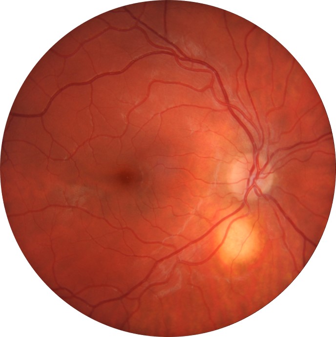
Funduscopy reveals a solitary, small yellow round lesion (about one disc diameter in size) most often located near the optic nerve and posterior to the equator. It may be associated with an orange halo.
OCT EDI imaging shows a smooth, dome shaped elevation within the sclera with partial or complete compression of the overlying choroidal vasculature. Rarely, subretinal fluid may be seen.
FAF mostly shows a homogenous mild hyper autofluorescence
B-scan ultrasound showed acoustic solidity
OCT angiography shows a clear choroidal flow overlying the lesion. The lesion itself is avascular.
Case Examples
Differential Diagnosis
References
Fung, AT, Waldstein, SM, Gal-Or, O, Pellegrini, M, Preziosa, C, Shields, JA, Lalane, R. (2020). Focal Scleral Nodule-A New Name for Solitary Idiopathic Choroiditis/Unifocal Helioid Choroiditis. Ophthalmology.