
Overview
Reactive RPE hyperplasia occurs secondary to ocular insults including trauma, intraocular inflammation, haemorrhage or retinal detachment. It is typically non progressive.
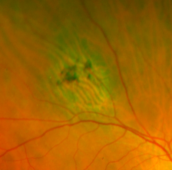
Funduscopic features include a well demarcated region of dark pigment with irregular borders. It may also be associated with chorioretinal atrophy.
OCT imaging shows RPE irregularity and hyperplasia with atrophy of the overlying retinal layers.
FAF shows corresponding hypoautofluorescence. There may also be areas of hyper autofluorescence which suggests RPE dysfunction.
Case Examples
-
Case 1: Unknown cause
A 70 year old Asian female with high myopia in the left eye and best corrected visual acuity of 6/7.5 in this eye. She does not recall any previous ocular trauma, injury or surgery.
-
Case 2: Unknown cause
A 58 year old Caucasian male with best corrected visual acuity of 6/6 (20/20) in the right eye.
-
Case 3: Chronic retinal detachment
An asymptomatic 51-year-old Asian male with high myopia and best corrected visual acuity of 6/30-1 (20/100-1) in the right eye and 6/6 (20/20) in the left eye.
-
Case 4: RPE hyperplasia associated with intrachoroidal cavitation
An asymptomatic 49 year old female with no signficant ocular or medical history. Visual acuity is 6/6 in this eye.
Differential Diagnosis
References
Ly, A. Nivison-Smith, L. Hennessy, M. Kalloniatis, M. (2015) Pigmented Lesions of the Retinal Pigment Epithelium, Optometry and Vision Science: Volume 92 - Issue 8
Shields JA, Shields CL. Tumors and Related Lesions of the Pigmented Epithelium. Asia Pac J Ophthalmol (Phila). 2017 Mar-Apr;6(2):215-223.