
Overview
Atypical CHRPE may be associated with conditions such as Gardner’s syndrome and Familial Adenomatous Polyposis (FAP). In this condition, multiple adenomatous polyps can be found in the colon and can progress to colorectal cancer. The presence of these lesions in the retina is often one of the earliest extraintestinal presentation of this genetic disorder and therefore accurate diagnosis is essential.
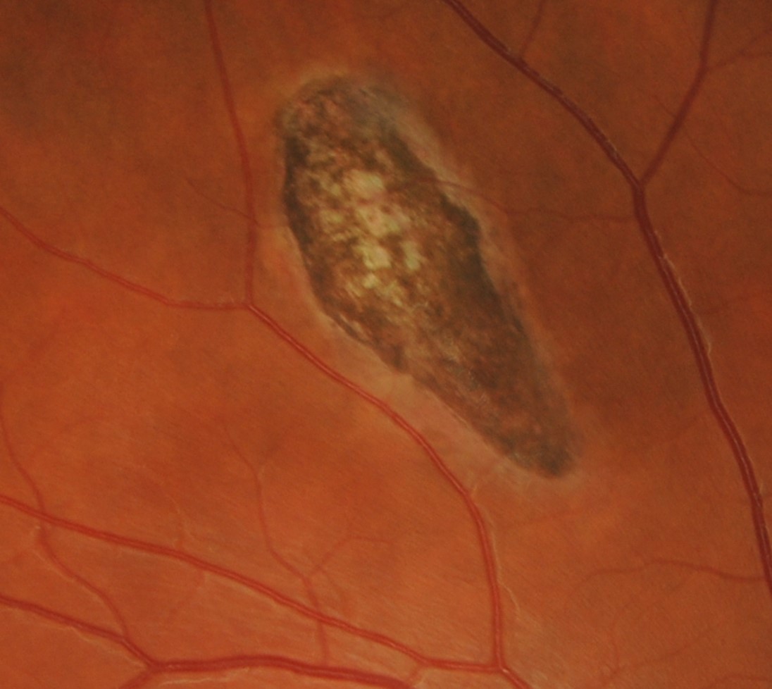
Atypical CHRPE are smaller and more ovoid than the typical CHRPE. They are often bilateral and are located haphazardly throughout the fundus. The surrounding border is often irregular and the edge of the lesion closest to the posterior pole takes the shape of a comma or fishtail with a depigmented tip. This is typically described as a "pisciform" lesion.
OCT imaging shows RPE thickening as well as outer retinal atrophy.
FAF shows corresponding hypo-autofluorescence with areas of iso/hyper autofluorescence
Case Examples
-
Case 1
A 31 year old Caucasian male with best corrected visual acuity of 6/6 (20/20) in the right eye. He reports good general health.
Differential Diagnosis
Congenital hypertrophy of the RPE (CHRPE)
Choroidal naevus
Toxoplasmosis
Grouped congenital hypertrophy of the RPE
Acquired RPE hyperplasia
Acute posterior multifocal placoid pigment epitheliopathy (APMPPE)
Multifocal choroiditis (MFC)
References
Ly, A. Nivison-Smith, L. Hennessy, M. Kalloniatis, M. (2015) Pigmented Lesions of the Retinal Pigment Epithelium, Optometry and Vision Science: Volume 92 - Issue 8
Nusliha et al., 2014. Congenital hypertrophy of retinal pigment epitheliu, (CHRPE) in patients with familial adenomatous polyposis (FAP): a polyposis registry experience
Raval et al., 2019. Multimodal imaging of congenital hypertrophy of the retinal pigment epithelium (CHRPE) lesions at different presentations
Wang et al., 2020. Multimodal imaging characteristics of congenital grouped hyper- and hypo-pigmented fundus lesions