
Diagnosis
Angle closure spectrum disease refers to a range of clinical presentations associated with narrow or occluded anterior chamber angles. Various grading scales exist for this disease spectrum, and in this resource we will use an adaptation of those proposed by Thomas et al. and the International Society for Geographical and Epidemiological Ophthalmology
1. Narrow but non-occludable angles: Narrow angles on primary gaze but angles are open on lens tilt indicating no iridotrabecular contact. It does not meet the criteria for PACS
3. Primary angle closure suspect (PACS) : PTM is not visible in 2 or more quadrants and/or iridotrabecular contact present. Normal IOP, no peripheral anterior synechiae and no optic nerve damage.
2. Primary angle closure (PAC): PACS plus elevated IOP and/or synechiae present but no optic neuropathy
3. Primary angle closure glaucoma (PACG): PACS or PAC with glaucomatous damage on visual fields or optic nerves. It does not require elevated pressures, symptoms or synechiae to be present.
Cases
-
Case 1: PAC
A 75 year old asymptomatic South American female with intraocular pressures of 37mmHg in each eye in the context of thick corneas (606µm RE, 614µm LE)
-
Case 2: PACG (RE) and PACS (LE)
A 77 year old asymptomatic Caucasian male with IOPs of 40mmHg in the right eye and 24mmHg in the left eye. These measurements were in the context of average corneal thicknesses of 558µm (RE) and 540µm (LE). He reports that his mother had high intraocular pressure, treated topically, however he was unsure if there was ever a formal diagnosis of glaucoma. His best corrected visual acuity was 6/6+ in each eye. There were no visible structures on gonioscopy in all quadrants in the right eye while the posterior trabecular meshwork was visible in the left eye in 2 quadrants only (nasal and inferior).
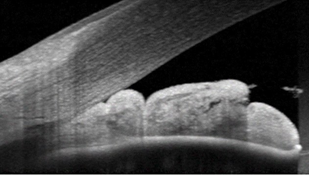
Anterior OCT (right eye)
More infoAnterior OCT (left eye)
More infoFundus photography (right and left eye)
More infoRed-free images (right and left eye)
More infoStereoscopic disc photo (right eye)
More infoStereoscopic disc photo (left eye)
More infoCirrus RNFL analysis
More infoCirrus Ganglion Cell Analysis
More info24-2 SITA-Standard visual field analysis
More info -
Case 3: PACS with synechiae
A 63 year old, hyperopic Asian male is asymptomatic and reports no significant family history. His intraocular pressures were 11mmHg in each eye in the context of average corneal thickness (543µm RE, 545µm LE). Gonioscopy showed angles in the both eyes to be open to the posterior trabecular meshwork inferiorly and Schwalbe's line in all other quadrants. Peripheral anterior synechiae was noted inferiorly in the left eye.
Goniophoto (inferior angle, left eye)
More infoPentacam Anterior Chamber Depth (right and left eye)
More infoDisc photos (right and left eye)
More infoFundus photographs (right and left eye)
More infoRed-free images (right and left eye)
More infoCIrrus RNFL analysis
More infoCirrus Ganglion Cell Analysis (GCA)
More info24-2 SITA-Standard Visual Field
More info
References
Devereux JG, Foster PJ, Baasanhu J, Uranchimeg D, Lee PS, Erdenbeleig T, Machin D, Johnson GJ, Alsbirk PH. Anterior chamber depth measurement as a screening tool for primary angle-closure glaucoma in an East Asian population. Arch Ophthalmol. 2000 Feb;118(2):257-63.
Foster PJ. Buhrmann R. Quigley HA et al. The definition and classification of glaucoma in prevalence surveys. Br J Ophthalmol 2002; 86: 238–242.
Phu, J. Hennessy, M. Spargo, M. Dance, S. Kalloniatis, M. (2020), A collaborative care pathway for patients with suspected angle closure glaucoma spectrum disease. Clin Exp Optom, 103: 212-219.
Phu J. Wong B. Lim T. Kalloniatis M. Assessment of angle closure spectrum disease as a continuum of change using gonioscopy and anterior segment optical coherence tomography. Ophthalmic Physiol Opt. 2020 Sep;40(5):617-631.
Thomas R, Walland MJ. Management algorithms for primary angle closure disease. Clin Exp Ophthalmol 2013; 41: 282–292.