
Overview
Angle recession occurs as a result of blunt ocular trauma. The force of the trauma displaces aqueous posteriorly and laterally, exerting a force on the iris root, causing a separation of the longitudinal and circular fibres of the cilliary body.
Angle recession due to blunt ocular trauma has been reported to be associated with secondary open angle glaucoma in 5-20% of cases with a higher risk associated with angle recession spanning more than 180 degrees.
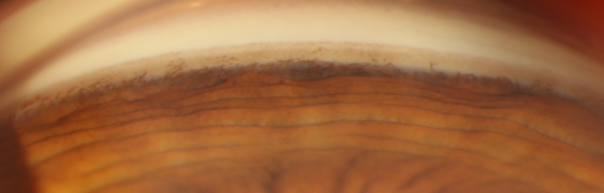
As discussed within the Gonioscopy page of this resource, angle recession is characterised by a focal widening of the ciliary body band on gonioscopy, and typically also increased trabecular meshwork pigmentation.
In the acute stages, traumatic forces may also cause damage to the trabecular meshwork, impeding aqueous outflow and leading to a subsequent increase in intraocular pressure. These same forces may also damage the anterior and/or posterior ciliary arteries, leading to hyphema (blood in the anterior chamber). The drainage of the red blood and inflammatory cells through the trabecular meshwork may further increase intraocular pressure.
Scarring and degeneration of the trabecular meshwork may cause increased intraocular pressure and subsequent glaucoma, even decades after the initial trauma.
Case Examples
-
Case 1: Ocular hypertension associated with angle recession
A 34 year old male with history of blunt trauma to his left eye 8 years ago. His visual acuity is 6/4.8- (20/16-) in each eye. His intraocular pressures were 21mmHg in the right eye and 27mmHg in the left in the context of thicker than average corneas (588µm OD and 590µm OS).
-
Case 2: Angle recession glaucoma
A 56 year old Caucasian male with a history of trauma to the right eye 16 years ago. 6 weeks after the trauma he underwent right cataract surgery. Prior to surgery, the patient had high myopia in both eyes. He reports no family history of glaucoma. His intraocular pressures were 22mmHg (OD) and 15mmHg (OS) in the context of average pachymetry values (568µm OD, 553µm OS).
Goniophotos (right eye)
More infoFundus photograph and red-free image (right eye)
More infoFundus photograph and red-free image (left eye)
More infoStereophoto of the right optic disc
More infoStereophoto of the right optic disc
More infoCirrus RNFL analysis
More infoCirrus Ganglion Cell Analysis (GCA)
More infoMatrix 24-2 FDT Threshold visual field analysis
More info
References
Kaufman JH, Tolpin DW. Glaucoma after traumatic angle recession a ten-year prospective study. Am J Ophthalmol. 1974;78
(4):648–654.
Ng, D.SC., Ching, R.HY. & Chan, C.WN. Angle-recession glaucoma: long-term clinical outcomes over a 10-year period in traumatic microhyphema. Int Ophthalmol 35, 107–113 (2015)
Senthil, S. Dangeti, D. Battula, M. Rao, HL. Garudadri, C. (2021): Trabeculectomy with Mitomycin-C in Post-Traumatic Angle Recession
Glaucoma in Phakic Eyes With no Prior Intraocular Intervention, Seminars in Ophthalmology