
Overview
Glaucoma is a progressive disease that needs to be monitored over time. Once progression is detected, the clinician may need to encourage adherence rates, modify or add to the treatment plan. Detecting progression can also help confirm the diagnosis of glaucoma by distinguishing true glaucoma from other mimicking disorders causing RNFL or visual field defects.
Glaucomatous progression can be identified using any, but preferably all, of the following methods:
1. Optic nerve head changes including neuroretinal rim loss, increase in RNFL defects or the presence of a disc haemorrhage
2. Progression on imaging: OCT progression analysis requires 2 baseline scans, from which later scans are compared to. OCT-based progression detection requires a significant reduction or downward trend in RNFL or GCIPL thickness.
3. Visual field progression: Indicated by the deepening or widening of existing defects, or the development of new defects.
Risk Factors for Progression
A 2012 study by Heijl et al. conlcuded that older age, higher mean IOP and IOP range were all associated with significantly faster progression.
The Early Manifest Glaucoma Trial (EMGT) showed that IOP lowering treatment in early glaucoma reduced the risk of progression risk in half. The risk of progression however is not equal between different types of glaucoma. The Normal Tension Glaucoma Study for example showed that while IOP lowering treatment will have a beneficial effect on most normal tension glaucoma patients, 20% will still continue to progress despite achieving IOP targets.
Case Presentations
-
Case 1: Structural and functional progression in a glaucoma suspect
A 47 year old Asian female with a family history of glaucoma in her brother presented for assessment.
Intraocular pressures (IOPs) were 22mmHg (OD) and 25mmHg (OS) in the context of average corneal thickness (568µm OD, 588µm OS). Gonioscopy showed angles open to the pigmented trabecular meshwork in all quadrants with no signs consistent with secondary glaucoma.The patient returned for follow-up 4 months after the initial consultation at which time IOPs were 19mmHg (OD) and 20mmHg (OS).
Fundus photographs at baseline and follow-up
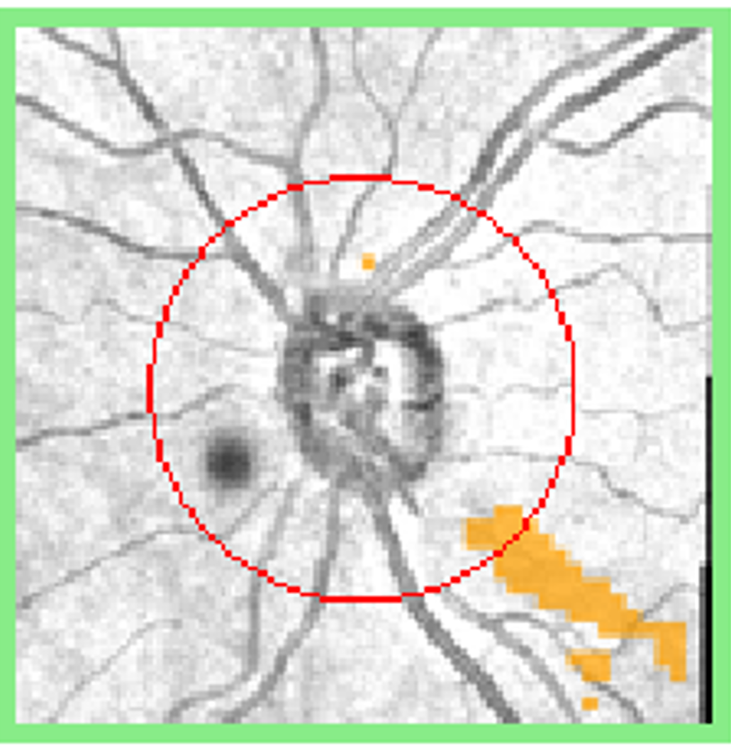
More infoRed-free images at baseline and follow up
More infoCirrus Guided Progression Analysis (extract)
More infoCirrus Ganglion Cell Analysis comparison (baseline and follow-up)
More info24-2 SITA-standard visual field analysis (baseline and follow-up)
More info -
Case 2: Disc haemorrhage and functional loss in a patient with glaucoma
A 74 year old Caucasian female with a history of thyroid disease who is being treated with Xalatan for bilateral glaucoma. At baseline, her intraocular pressure (IOP) in the right eye was 17mmHg in the context of average corneal thickness (562µm). At follow-up 6 months later, her IOP was the same.
-
Case 3: Disc haemorrhage and structural loss in a glaucoma suspect
A 54 year old male with a family history of glaucoma (mother). His intraocular pressure (IOP) is 24mmHg in the context of average corneal thickness (515µm OD and 515µm OS). Angles are open to the ciliary body band and there is no signs suggestive of secondary glaucoma.
-
Case 4: Progression of central field loss in a patient with glaucoma
A 52 year old myopic Asian male with a history of smoking and hypertension. He also reports a family history of glaucoma (mother). He was diagnosed 1 year prior with normal tension glaucoma and has had SLT 6 months previous. His intraocular pressure in the right eye is 15mmHg in the context of average corneal thickness (pachymetry 525µm). Gonioscopy shows the angle open to the ciliary body band in all quadrants and there are no signs suggestive of secondary glaucoma.
References
Anderson, DR. (2003) Collaborative Normal Tension Glaucoma Study, Current Opinion in Ophthalmology: April 2003 - Volume 14 - Issue 2 - p 86-90
Heijl A. Buchholz, P. Norrgren G. and Bengtsson B. (2013), Rates of visual field progression in clinical glaucoma care. Acta Ophthalmologica, 91: 406-412.
Leskea, M. Heijl A. Hyman, L. Bengtsson, B, Komaroff, E. (2004) Factors for progression and glaucoma treatment: The Early Manifest Glaucoma Trial, Current Opinion in Ophthalmology: Volume 15 - Issue 2 - p 102-106