
Overview
Pseudoexfoliation syndrome is associated with the presence of white-grey, fibrillar, pseudoexfoliative material visible in the anterior segment of the eye. This material may be found on any structure, but is most visible on the anterior lens surface following pupil dilation, appearing as a white double ring or "bulls-eye" pattern with the inner ring located at the resting pupil border.
Other ocular structures where this pseudoexfoliative matieral may be visible include the pupil margin and anterior surface of the iris. The material may also be deposited in the anterior chamber angle, and a progressive buildup over time can lead to progressively increasing intraocular pressure and pseudoexfoliative glaucoma. Glaucoma associated with pseudoexfoliation syndrome is typically bilateral but asymmetric, increases with age, and has faster rates of progression than primary open angle glaucoma.
Additional effects of this condition on the structures of the eye include fragility of the lens zonules, leading to an increased risk of lens subluxation and increased complications from cataract surgery. Pseudoexfoliation syndrome is also associated with disruption of the blood-aqueous barrier, a lower number of corneal endothelial cells and poor or absent pupillary dilation.
Pseudoexfoliation syndrome is a systemic condition rather than an isolated ocular condition. The extracellular matrix is affected not only in the eye, but also in the body's other visceral organs, resulting in the deposition of pseudoexfoliative material around blood vessels in connective tissue.
Imaging Characteristics
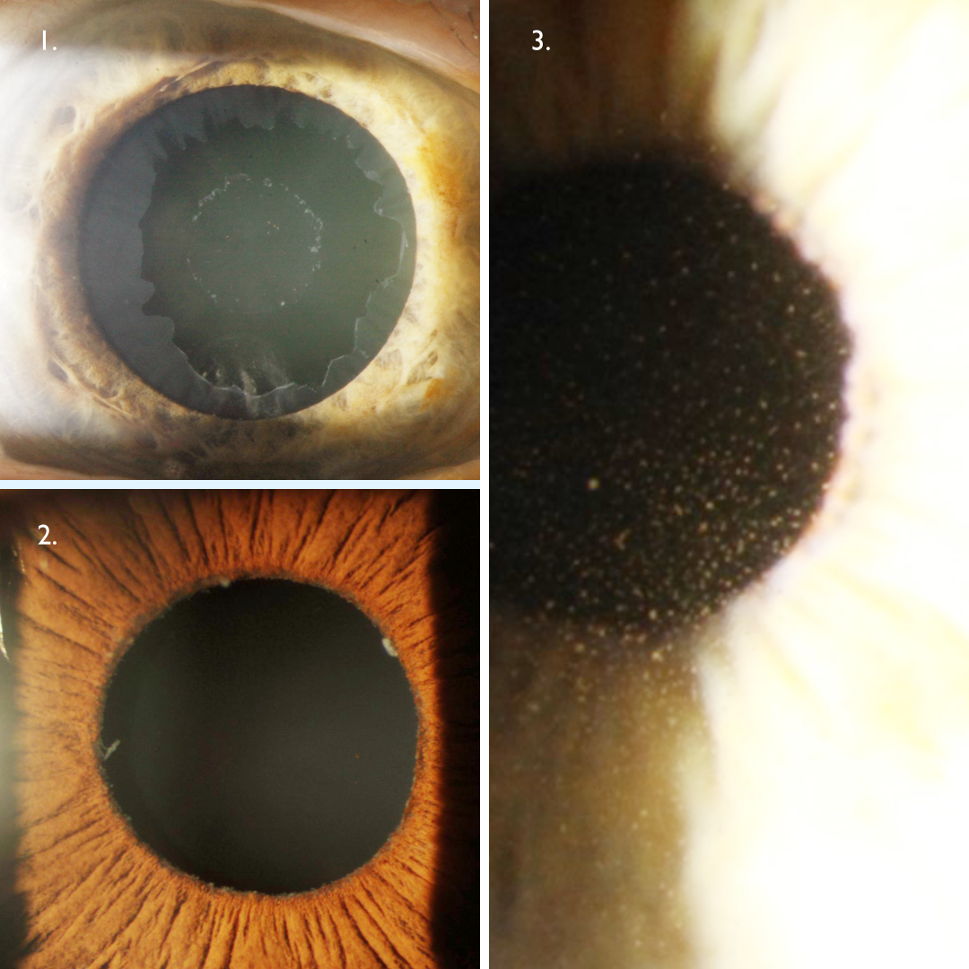
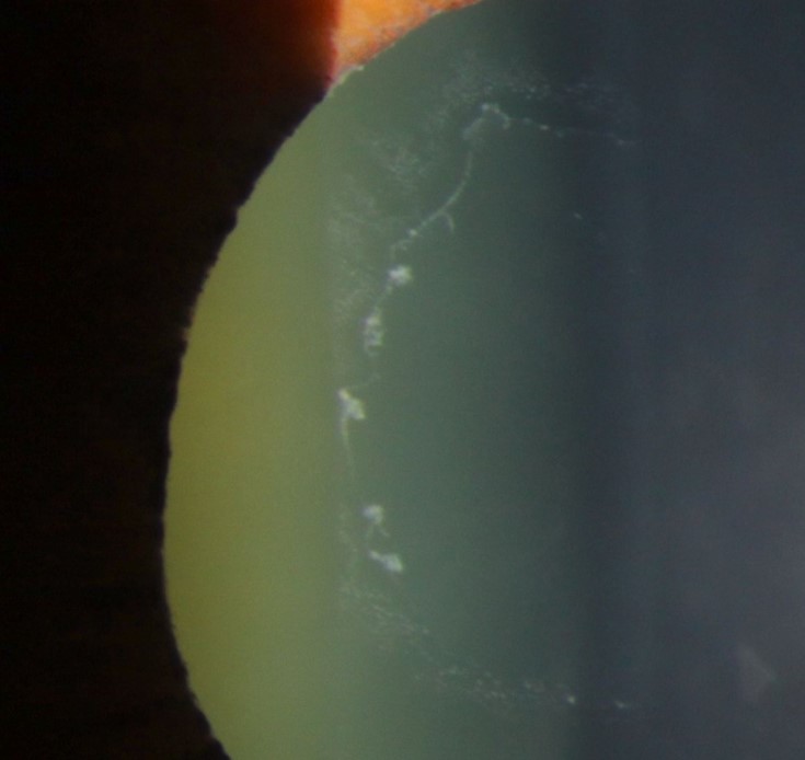
Image 1 shows pseudoexfolative, white material on the anterior lens with a dilated pupil. Note the classical bull’s eye distribution; with an inner ring at the resting pupil border surrounded by a clear zone.
Image 2 shows pseudoexfoliative material on the pupil margin. This is often associated with loss of pupillary ruff as you can see in the photograph. Retroillumination can be particularly useful to observe transillumination defects at the pupil margin.
Image 3 illustrates extensive endothelial pigment deposition that is associated with PXF syndrome.
Gonioscopy shows increased, patchy pigmentation of the trabecular meshwork and Schwalbe's line.
Case Examples
-
Case 1: Pseudoexfoliation syndrome
A 58 year old Middle Eastern female with intraocular pressures of 15mmHg in each eye in the context of thin corneas (494µm OD, 496µm OS). The right eye showed no anomalies, so only the left eye will be discussed in this case.
-
Case 2: Pseudoexfoliation syndrome
A 76 year old female with intraocular pressures of 19mmHg in each eye at presentation, in the context of average corneal thicknesses (559µm OD, 561µm OS). This patient has been followed at CFEH for 7 years since this initial visit with IOP ranging from 14mmHg to 20mmHg over this time.
Anterior eye photographs
More infoAnterior eye photograph using retroillumination
More infoGoniophoto (right superior angle)
More infoFundus photography (right and left eye)
More infoCirrus RNFL analysis
More infoCirrus Ganglion Cell Analysis
More infoCirrus Guided Progression Analysis (right eye)
More infoCirrus Guided Progression Analysis (left eye)
More info -
Case 3: Pseudoexfoliation glaucoma
A 68 year old Indian female with intraocular pressures of 19mmHg (OD) and 24mmHg (OS) in the context of average corneal thicknesses (511µm OD, 516µm OS). She has type 2 diabetes, hypertension, hyperlipidaemia and is a migraine sufferer.
Gonioscopy showed the angles open to the ciliary body band in all quadrants with increased, non-homogenous pigmentation of the trabecular meshwork inferiorly.
Differential Diagnosis
References
Elhawy E. Kamthan G. Dong CQ. Danias J. (2012) Pseudoexfoliation syndrome, a systemic disorder with ocular manifestations. Hum Genomics. 2012;6(1):22.
Grødum K. Heijl A. Bengtsson B. (2005) Risk of glaucoma in ocular hypertension with and without pseudoexfoliation. Ophthalmology. 2005;112(3):386–390.
Plateroti P. Plateroti AM. Abdolrahimzadeh S. Scuderi G. (2015) Pseudoexfoliation Syndrome and Pseudoexfoliation Glaucoma: A Review of the Literature with Updates on Surgical Management. J Ophthalmol. 2015:370371