
The Normal Macula
The macula is a horizontally oval area approximately 15 degrees temporal to the optic nerve and is the point of the eye where images from the central vision are focussed by the optics of the eye. For this reason, the macula has the highest concentration of photoreceptors, allowing us to perceive high resolution central visual acuity.
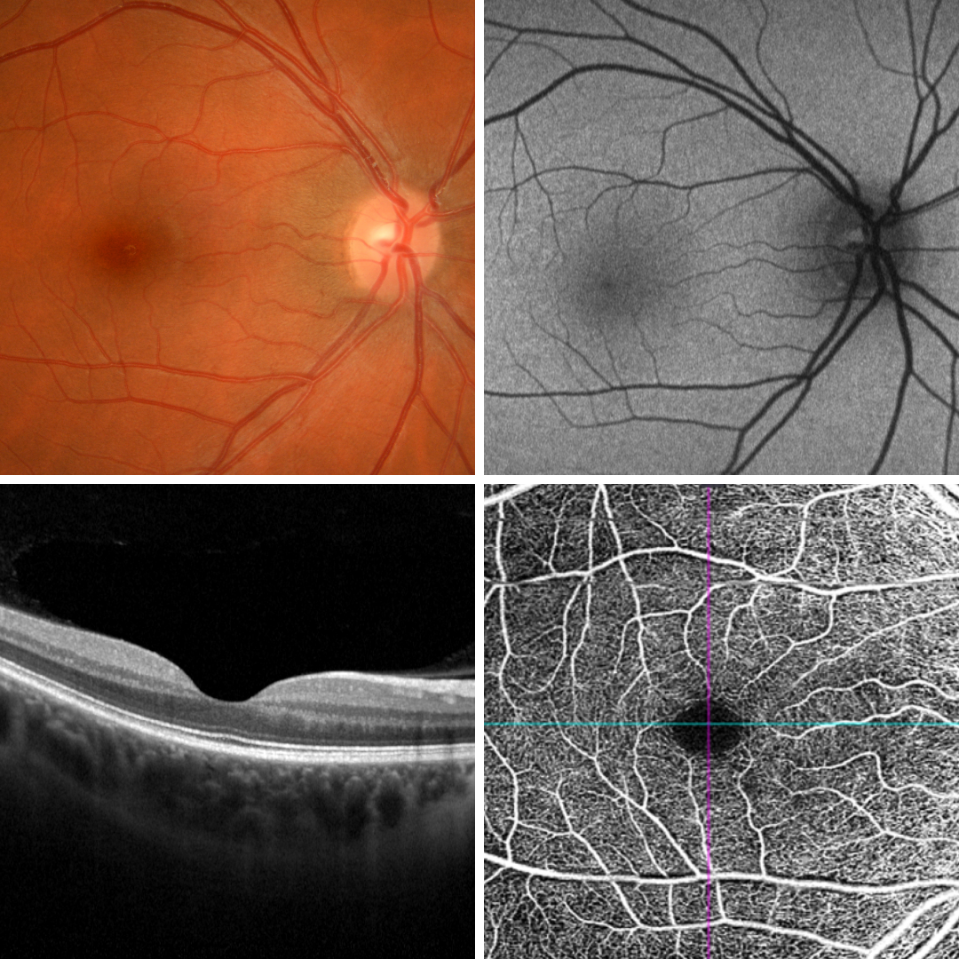
On funduscopy, the macula appears darker in colour than the surrounding retina, and it appears slightly hypo-autofluorescent when compared with the surrounding retina on fundus autofluorescence imaging. At the centre of the macula is the fovea which shows a “foveal reflex” on funduscopy.
The fovea is approximately 0.35mm in diameter and is the point at which images from the centre of vision are imaged. The concentration of cone photoreceptors at the fovea is much greater than anywhere else in the retina.
The fovea is devoid of the inner retinal layers and has a characteristic shape as seen on the OCT provided. There are also no retinal blood vessels at the fovea as seen on the OCT angiography image. The foveal avascular zone (FAZ) is approximately 0.6,, in diameter although this varies with age and can enlarge in certain diseases (eg diabetic retinopathy).
Damage to the macula can have a significant impact on central vision and the early detection of macular disease can, in many cases, help prevent or reduce damage to this critical part of the eye.