
Overview
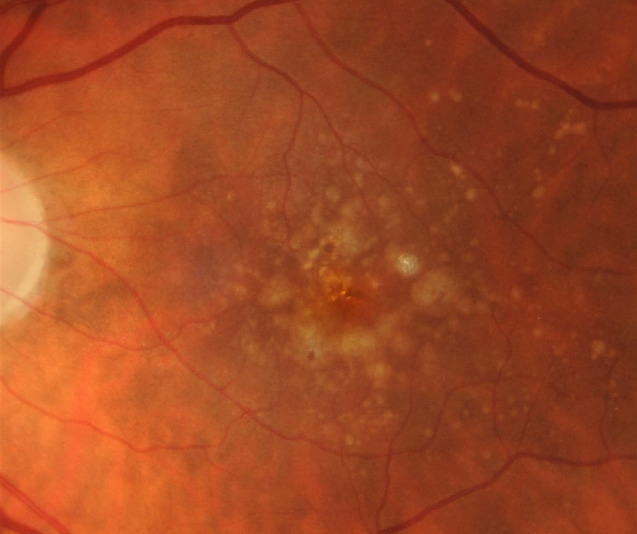
In AMD, pigmentary abnormalities refer to the presence of hyper-pigmentation or hypo-pigmentation within two disc diameters of the fovea with drusen 63μm or more in diameter. It is a feature of at least intermediate AMD and is associated with a higher risk of progression to late AMD.
Hypo-pigmentary changes appear clinically as depigmented areas due to a reduction in pigment content, thinning or loss of the RPE. They can be irregular in shape with poorly defined borders.
Hyper-pigmentary changes appear as gray or black pigment deposits. On OCT imaging, hyper-pigmentary abnormalities may present as hyper-reflective foci within the retina.