
Overview
Described here is a simple way to estimate drusen size using anatomical cues, a skill that is necessary for the accurate classification of AMD.
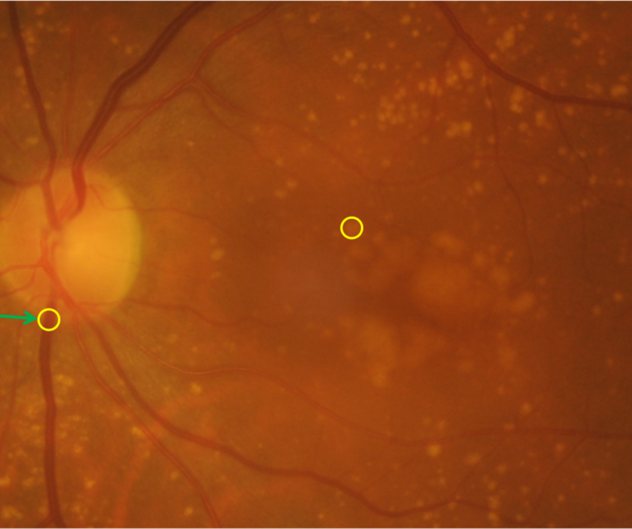
The diameter of the central retinal vein at the optic disc margin is approximately 125µm and this is used to give us a point of comparison.
- Small drusen are less than 63µm - that is, less than half the vein diameter as it crosses the edge of the optic disc.
- Medium drusen are between 63µm and 125µm - that is, more than half but less than the full diameter of the vein
- Large drusen are larger than 125µm - that is, they have a diameter larger than the vein
Case Examples
-
Small drusen
Small drusen (drupelets) are smaller than 63µm in diameter.
-
Medium drusen
Medium drusen are between 63µm and 125µm in diameter.
-
Large drusen
Large drusen have a diameter greater than 125µm.