
Overview
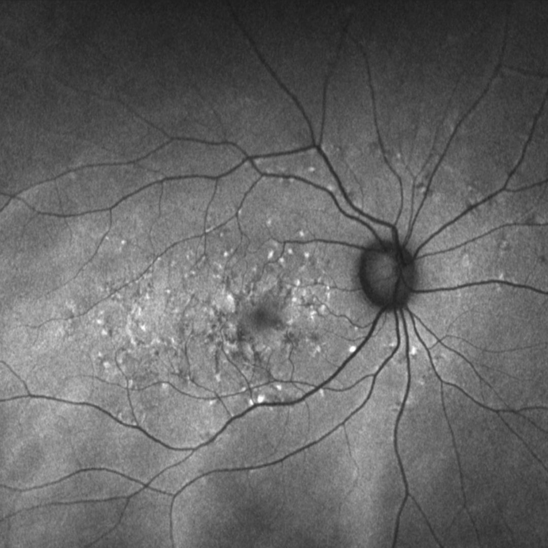
Similar to Stargardt disease, multifocal pattern dystrophy presents with irregular yellow flecks within the posterior pole. These flecks are initially hyper-autofluorescent but can change over time. OCT shows disturbance and abnormalities in the photoreceptor outer segment-RPE level.
The two conditions may be difficult to differentiate. Multifocal pattern dystrophy can be differentiated from Stargardt disease based on its autosomal dominant inheritance pattern (whereas Stargardt disease is typically autosomal recessive), later age of onset, a comparatively better visual prognosis, and the absence of dark choroid on fluorescein angiography.
Case Example
-
Case 1
A 57-year-old Middle Eastern female with best-corrected acuities of 6/6+ (20/20+) in each eye.
Fundus photograph and red free image (right eye)
More infoFundus photograph and red free image (left eye)
More infoOptomap and fundus autofluorescence image (right eye)
More infoOptomap and fundus autofluorescence image (left eye)
More infoSpectralis OCT volume and line scans (right macula)
More infoSpectralis OCT volume and line scans (left macula)
More info