
Overview
Stargardt disease is one of the most common juvenile-onset macular dystrophies affecting 1 in 10,000 persons, associated with autosomal recessive inheritance of a mutation in the ABCA4 gene.
The condition features progressive central vision loss with multifocal yellow-white fundus flecks and atrophic macular lesions.
Although Stargardt disease is typically diagnosed within the first two decades of life, late-onset have also been reported and the latter is associated with foveal sparing of atrophic changes and better visual prognosis.
Due to the accumulation of lipofuscin, the fundus flecks in Stargardt disease display intense hyper-autofluorescence during their active stage. However, the autofluorescence signal reduces as the flecks resorb and RPE atrophy develops.
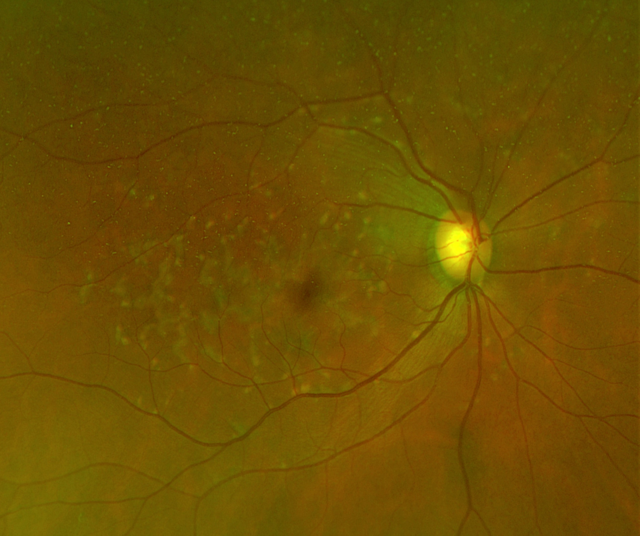
The other characteristic clinical feature of Stargardt disease is peripapillary sparing where an annulus of normal retina tissue free from flecks and RPE atrophy can be found surrounding the optic nerve head.
Dark choroid sign in fluorescein angiography can be found in over 80% of Stargardt patients and is useful to differentiate Stargardt disease from multifocal pattern dystrophy simulating Stargardt.
Fundus flavimaculatus shares a similar phenotype and link to the same genetic mutation with Stargardt disease and is considered to be the same disease.
Case Examples
-
Case 1: Stargardt Disease (foveal sparing)
A 46-year-old Caucasian male with best-corrected visual acuities of 6/9.5 (20/33) in the right eye and 6/7.5 (20/25) in the left.
Colour fundus photograph and red free image (right eye)
More infoFundus photography and red free image (left eye)
More infoOptomap and fundus autofluorescence images (right and left eye)
More infoSpectralis OCT series of line scans (right and left macula)
More infoSpectralis OCT line scans
More infoSpectralis OCT macular line scans (right - top and left - bottom)
More info -
Case 2: Stargardt Disease
A 22-year-old Caucasian male with best-corrected visual acuity of 6/15 in the right and 6/12 in the left. Monocular colour vision testing showed a Deutan defect in each eye.
-
Case 3 - Stargardt Disease
A 46 yo Caucasian male referred for suspected retinal dystrophy. Noted 4-6 months of worsening distance vision, particularly at night time. He had not had an eye examination for 20 years prior.
Colour fundus photographs
More infoOptomap wide field images and fundus autofluoresence (FAF) images
More infoCirrus OCT macular cube (right eye)
More infoCirrus OCT macular cube (left eye)
More infoCirrus OCT Angiography (right macula, 6x6mm ORCC)
More infoCirrus OCT Angiography (left macula, 6x6mm ORCC)
More info -
Case 4 - Fundus Flavimaculatus
A 59 year old Caucasian female with best corrected acuities of 6/6 (20/20) in each eye.
Differential diagnosis
References
Cideciyan AV, Swider M, Aleman TS et al. (2005) ABCA4-associated retinal degenerations spare structure and function of the human parapapillary retina. Invest Ophthalmol Vis Sci 46: 4739-4746.
Fishman GA, Farber M, Patel BS (1987) et al. Visual acuity loss in patients with Stargardt's macular dystrophy. Ophthalmology 94: 809-814.
Georgiou M, Kane T, Tanna P et al. (2020) Prospective Cohort Study of Childhood-Onset Stargardt Disease: Fundus Autofluorescence Imaging, Progression, Comparison with Adult-Onset Disease, and Disease Symmetry. American Journal of Ophthalmology 211: 159-175.
Lambertus S, van Huet RA, Bax NM et al. (2015) Early-onset stargardt disease: phenotypic and genotypic characteristics. Ophthalmology 122: 335-344.
Michaelides M, Hunt DM, Moore AT. (2003) The genetics of inherited macular dystrophies. Journal of medical genetics 40: 641-650.
Rotenstreich Y, Fishman GA, Anderson RJ. (2003) Visual acuity loss and clinical observations in a large series of patients with Stargardt disease. Ophthalmology 110: 1151-1158.
Saksens NT, Fleckenstein M, Schmitz-Valckenberg S et al. (2014) Macular dystrophies mimicking age-related macular degeneration. Prog Retin Eye Res 39: 23-57.
Stargardt K. (1909) Über familiäre, progressive Degeneration in der Maculagegend des Auges. Albrecht von Graefes Archiv für Ophthalmologie 71: 534-550.
Van Huet RA, Bax NM, Westeneng-Van Haaften SC et al. (2014) Foveal sparing in Stargardt disease. Invest Ophthalmol Vis Sci 55: 7467-7478.
Westeneng-van Haaften SC, Boon CJ, Cremers FP et al. (2012) Clinical and genetic characteristics of late-onset Stargardt's disease. Ophthalmology 119: 1199-1210.