
Overview
Autosomal dominant drusen are also referred to as Malattia Leventinese or Doyne’s honeycomb retinal dystrophy. It is caused by a mutation in the EFEMP1 gene and is associated with an autosomal dominant inheritance pattern.
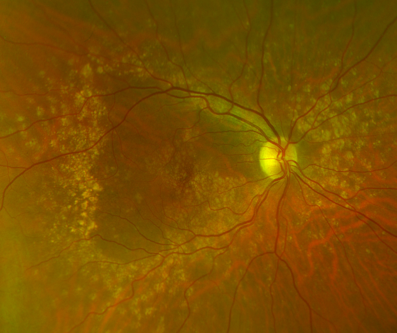
It presents with characteristic early-onset (before 50) drusen at the macula, posterior pole, and/or abutting optic nerve head.
The onset is in the 3rd to 4th decade of life. Patients may be asymptomatic at first but can develop reduced vision, metamorphosias or scotomas later in life. There is a risk of developing geographic atrophy and choroidal neovascularisation.
Fundus photography and wide field imaging typically show small drusen in a radial distribution temporal to the macula, large drusen at the posterior pole, and/or drusen nasal to the optic nerve head. Drusen distribution is relatively symmetrical between the eyes.
Case Examples
-
Case 1
A 41-year-old female with best-corrected visual acuity of 6/6 (20/20) in the right eye and 6/7.5 (20/25) in the left.
Fundus photography (right and left eye)
More infoOptomap (1), red separation (2) and green separation (3) images - right eye
More infoOptomap (1), red separation (2) and green separation (3) images - left eye
More infoSpectralis OCT volume scan and macula line scan (right eye)
More infoSpectralis OCT volume scan and macula line scan (left eye)
More infoFundus autofluoresence images (right and left eye)
More info -
Case 2
A 42-year-old Caucasian female with best-corrected visual acuity of 6/4.8- (20/15-) in each eye.
Fundus photography (right and left eye)
More infoRed-free images (right and left eye)
More infoWidefield and fundus autofluoresence images (right and left eye)
More infoSpectralis OCT line scans (right and left macula)
More infoSpectralis OCT line scans taken through areas of drusen (midperipeheral retina - right and left eye)
More info
Differentials
References
Kumar, N., Balyan, M., & Bansal, R. (2020). Multimodal imaging in familial dominant drusen. Indian journal of ophthalmology, 68(10), 2266.
Oishi, A., Oishi, M., Miyata, M., Hirashima,T., Hasegawa, T., Numa, S., Tsujikawa, A (2018) Multimodal Imaging for Differential Diagnosis of Bietti Crystalline Dystrophy, Ophthalmology Retina, Volume 2, Issue 10, Pages 1071-1077.
Stefko ST, Zhang K, Gorin MB, Traboulsi EI. (2000) Clinical spectrum of chromosome 6-linked autosomal dominant drusen and macular degeneration. Am J Ophthalmol. Aug;130(2):203-8.