
Overview
Best vitelliform macular dystrophy, also known as Best disease, is a clinically heterogeneous disease mostly associated with a mutation in BEST1 gene and has an autosomal dominant pattern of inheritance.
Best disease usually presents in the first decade of life. Visual acuity is minimally affected in the early stages of the disease but symptoms can start to develop in the 3rd to 4th decade.
Best disease can be classified into different stages based on fundus examination, although the stages do not always occur consecutively:
1. Previtelliform stage: Fundus may be normal or show mild pigmentary changes, but OCT shows increased thickness and reflectivity in the interdigitation zone and EOG is abnormal.
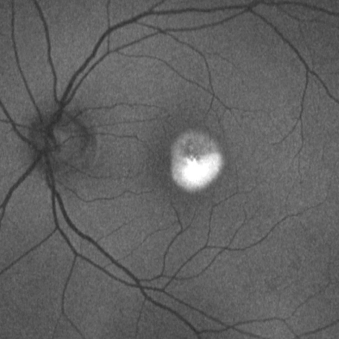
2. Vitelliform stage: A classic "egg yolk lesion" is visible in the fundus, usually ½-2 disc diameter in size. On OCT, the lesion is seen to be sub-retinal in location and vision is unaffected at this stage.
3. Pseudohypopyon stage: The vitelliform lesion starts to regress and the heavier vitelliform material gravitates down the subretinal space.
4. Vitelliruptive stage: The macula shows a "scrambled egg" appearance as the dispersion of the vitelliform material occurs.
5. Atrophic stage: Complete resorption of the vitelliform material occurs and chorioretinal atrophy ensues. This stage can resemble the geographic atrophy in AMD and vision can be greatly reduced.
6. Cicatricial stage: This features the development of choroidal neovascularization and can occur at any stage of Best disease.
Diagnosis of Best disease is based on the clinical presentation, family history, and a decreased electrooculogram (EOG) light peak:dark trough ratio (previously known as Arden ratio) and normal full-field electroretinogram.
Case Examples
-
Case 1: Vitelliform stage
A 54-year-old Caucasian male with visual acuity of 6/6 (20/20) in each eye.
Colour fundus photograph and red-free image (right eye)
More infoColour fundus photograph and red-free image (left eye)
More infoOptomap widefield and fundus autofluorescence images (right - top, left- bottom)
More infoSpectralis OCT volume scan and macula line scan (right eye)
More infoSpectralis OCT volume scan and macular line scan (left eye)
More info -
Case 2: Pseudohypopyon
A 52-year-old Middle Eastern male with best-corrected acuity of 6/12 (20/40) in his left eye.
-
Case 3: Vitelliruptive stage
A 48-year-old Caucasian male with best-corrected visual acuity of 6/38 (20/130) in his right eye. He also has strabismic amblyopia in this eye.
-
Case 4: Cicatrical Stage
A 14-year-old Middle Eastern male with best-corrected acuity of 6/15 (20/50) in the left eye.