
Overview
Adult-onset foveomacular vitelliform dystrophy is characterised by bilateral subfoveal yellowish subretinal deposits. These deposits typically span one third of a disc diameter, and show intense hyper-autofluorescence. In some cases, multifocal vitelliform lesions are present.
Over time, pigmentary changes, progressive atrophy or choroidal neovascularisation may develop.
Adult-onset foveomacular vitelliform dystrophy can be classified into vitelliform, pseudohypopyon, vitelliruptive, and atrophic stages. Visual prognosis is usually favourable but vision loss can occur from choroidal neovascularisation in later stages.
Case Examples
-
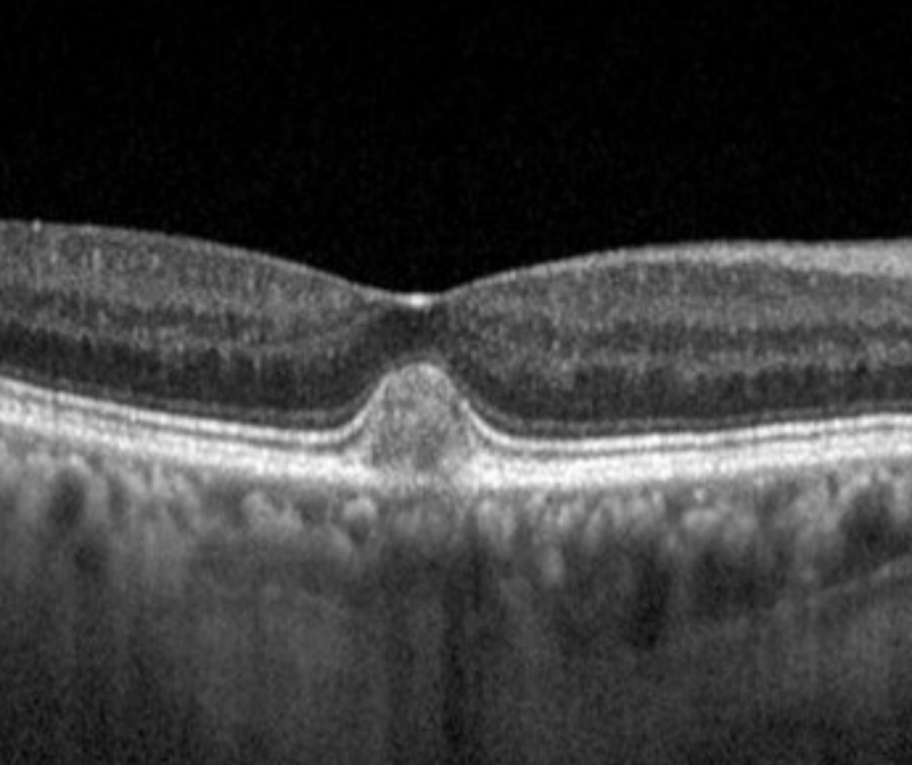
Case 1: Adult-onset foveomacular vitelliform dystrophy (vitelliform stage)
A 72-year-old Caucasian male with visual acuity of 6/7.5 (20/25) in each eye.
Differential diagnosis
Best disease
Polypoidal choroidal vasculopathy (PCV)
Neovascular AMD
Myopic choroidal neovascularisation
Acute central serous choroiretinopathy (CSCR)
Retinal toxicity (Deferoxamine retinopathy)
Acute exudative polymorphous vitelliform maculopathy.
Additional differentials include acquired vitelliform lesions secondary to age-related macular degeneration (AMD), vitreomacular traction (VMT), pseudoxanthoma elasticum maculopathy and mitochondrial retinal dystrophy,