
Overview
A macular coloboma is a rare variant of a typical coloboma (described elsewhere in this resource - link provided below). A coloboma occurs due to the incomplete closure of the embryonic fissure during foetal development.
Macular colobomas are often bilateral and symmetric between the two eyes. They most commonly have a genetic cause (possibly associated with other systemic or ocular abnormalities), however it is believed they can also arise secondary to an intrauterine inflammation.
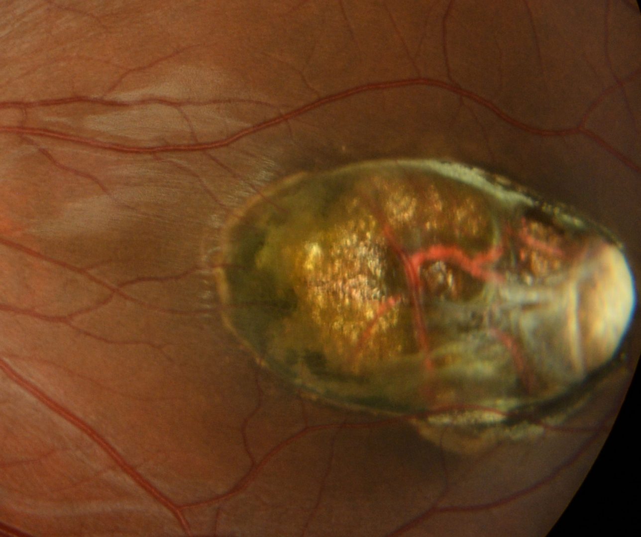
Clinically they present as large oval lesions located in the central posterior pole. They are excavations associated with sharply defined borders and variable pigmentation is typically associated.
Case Example
-
Case 1: Unilateral macular coloboma
A 20 year old Middle Eastern male with a long-standing history of poor vision in his left eye. Best corrected visual acuity was 6/48- (20/160-). He reports good general health and takes no medications.
Differential Diagnosis
References
Parmeggiani, F., Milan, E., Costagliola, C. et al. Macular coloboma in siblings affected by different phenotypes of retinitis pigmentosa. Eye 18, 421–428 (2004).
Varghese, M., Kavalakatt, J. A., Pandey, S., & Kolath, J. J. (2016). Macular coloboma. Oman journal of ophthalmology, 9(1), 67–68.