
Overview
Solar retinopathy is a photochemical retinal injury caused by either direct or indirect viewing of the sun. The risk of damage is higher in patients taking drugs causing photosensitivity such as Tetracycline and Psoralens, while it is reduced in patients with darkly pigmented retinae and those with high refractive errors.
Symptoms of solar retinopathy include decreased vision (in the range 6/7.5 – 6/30) with associated central scotomas. Metamorhopsia, dyschromatopsia, micropsia and headaches occurring within hours of exposure are also commonly experienced.
There is no effective treatment for solar retinopathy. In many cases, visual acuity may recover to normal, however some patients have residual small central or paracentral scotomas.
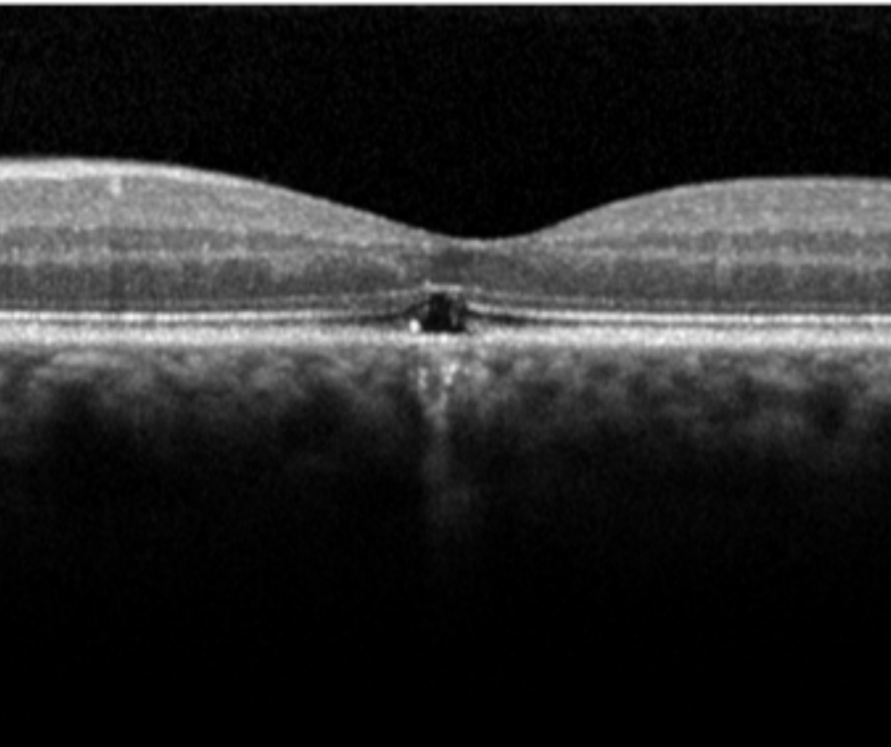
Fundus findings initially include a yellow-white spot in the fovea, changing to a red spot with a pigmented halo over several days. On fundus autofluoresence, solar retinopathy appears as hypo-autofluorescence at the fovea surrounded by an irregular ring of hyper-autofluorescence. OCT imaging typically shows disruption of the inner and/or outer segments of the photoreceptor layers, either with or without RPE defects.
The literature indicates that defects of the inner photoreceptor segment are associated with worse VA than defects in either the outer segment or RPE. There may also be a transient increase in foveal reflectivity and reduced reflectivity of the RPE.
Case Examples
-
Case 1
A 48 year old Caucasian male with best corrected acuity of 6/12 (20/40) in the right eye and 6/15 (20/50) in the left. He reports a history of working with lasers.
-
Case 2
A 48 year old Caucasian male with a history of extended sun gazing, since which time he has noticed reduced vision in both eyes. His best corrected visual acuity is 6/12- (20/40-) in the right eye and 6/15 (20/50) in the left.
Differential Diagnosis
References
Jain, A. Desai, RU. Charalel, RA. Quiram, P. Yannuzzi, L. Sarraf, D. (2009) Solar Retinopathy Retina: Volume 29 - Issue 9 - p 1340-1345