
Overview
Dome-shaped macula refers to a convex elevation of the macula ( including the retina, RPE and choroid), usually within an area of a posterior staphyloma. It is a diagnosis most easily made with a vertical OCT line scan through the macula. Chronic sub-retinal fluid may be present, in which case the condition is termed dome-shaped maculopathy.
Dome-shaped maculopathy may be associated with reduced vision and metamorphopsia, or patients may be asymptomatic.
Case Examples
-
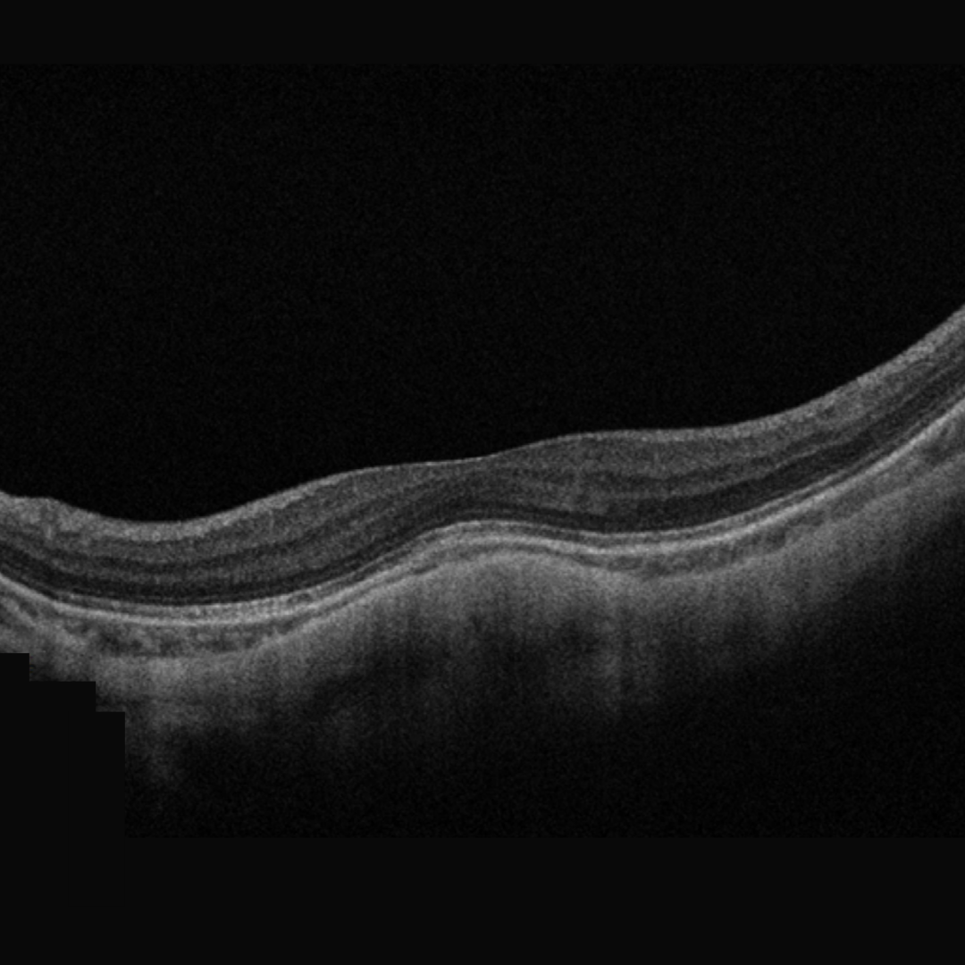
Case 1: Dome-shaped macula with no sub-retinal fluid
A 61 year old Asian male with prescription of -17.25 DS in the right eye and best corrected visual acuity of 6/12- (20/40-).
-
Case 2: Dome-shaped macula with no sub-retinal fluid
A 64 year old Middle Eastern female with best corrected visual acuity of 6/15 in the right eye. She had cataract surgery five years previously. Her pre-surgery refraction is unknown but she reports it to be high myopia that was treated in the right eye with a Thompson's sling several years previous.
Differential Diagnosis
References
Lee, GW., Kim, JH., Kang, SW. et al. (2020) Structural profile of dome-shaped macula in degenerative myopia and its association with macular disorders. BMC Ophthalmol 20, 202.
Ng, D., Cheung, C., Luk, F. et al. (2016) Advances of optical coherence tomography in myopia and pathologic myopia. Eye 30, 901–916
Yasushi, I. (2017) Overview of the complications of high myopia, Retina: Volume 37 - Issue 12 - p 2347-2351