
Overview
A Föster-Fuchs spot is a pigmented scar of regressed myopic CNV.
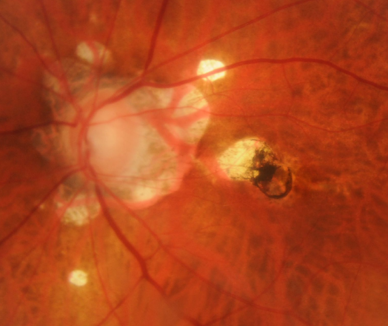
On Fundus photos, Förster-Fuchs spots appear as raised, round pigmented lesions of RPE proliferation adjacent to areas of chorioretinal atrophy consistent with regressed choroidal neovascularization.
OCT typically shows RPE hyperplasia with adjacent chorioretinal atrophy and a distinct increase in light transmission through this area.
Fundus autofluorescence imaging often shows a focal area of hypoautofluorescence due to signal blockage from the RPE hyperplasia and chorioretinal atrophy
Case Examples
-
Case 1
A 65 year old Middle Eastern female with refraction of approximately -10.00D prior to cataract surgery. Best corrected visual acuity is 6/6- (20/20-) and she has a previous history of having 2 anti-VEGF injections in her left eye.
Differential diagnosis
References
Ng, D., Cheung, C., Luk, F. et al. (2016) Advances of optical coherence tomography in myopia and pathologic myopia. Eye 30, 901–916
Yasushi, I. (2017) Overview of the complications of high myopia, Retina: Volume 37 - Issue 12 - p 2347-2351
Ohno-Matsui, K., Ikuno, Y., Lai, TTI., Cheung, CMG (2018) Diagnosis and treatment guideline for myopic choroidal neovascularization due to pathologic myopia. Progress in Retinal and Eye Research, Volume 63, Pages 92-106,
Milani, P., Massacesi, A., Moschini, S., Setaccioli, M., Bulone, E., Tremolada, G., Ciaccia, S., Mantovani, E., Morale, D., & Bergamini, F. (2016). Multimodal imaging and diagnosis of myopic choroidal neovascularization in Caucasians. Clinical ophthalmology (Auckland, N.Z.), 10, 1749–1757.