
Overview
Lacquer cracks present as ruptures in Bruch’s membrane caused by stretching of the posterior fundus with axial elongation. They may radiate from the disc, or through and around the macula. Although thought to be associated with a staphyloma, research data suggests that there is no obvious correlation with either axial length or refraction.
Lacquer cracks present as fine irregular, yellow white lines in the posterior fundus.
OCT scans may show a discontinuity in the RPE with increased light penetration into posterior tissue. Lacquer cracks are often best visualised using infra-red imaging.
Fundus autofluorescene imaging can show a hypo autofluorescent pattern corresponding to the lacquer crack. Infrared imaging may be superior to FAF in the detection rate and shows hyper reflective lines.
Lacquer cracks typically progress over time, either in number or extent but they usually do not affect vision unless macula haemorrhage or choroidal neovascularization occurs.
Case Examples
-
Case 1: Lacquer crack
A 60 year old Asian female with refraction:
Right eye: -6.00 / -1.25 x 90 VA 6/6 (20/20)
Left eye: -5.00 / -1.75 x 90 VA 6/6 (20/20) -
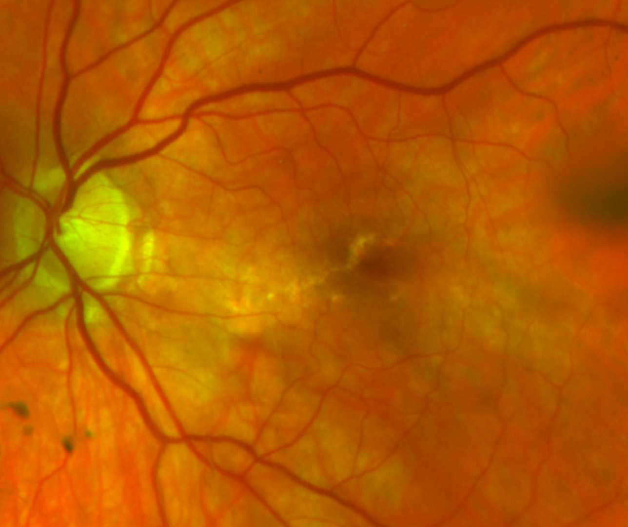
Case 2: Lacquer crack at the macula
A 70 year old Caucasian male with refraction -3.75 / -1.25 x 70 VA 6/6- (20/20-)
-
Case 3: Lacquer crack
A 55 year old Asian female presented for examination. Pinhole visual acuity was 6/7.5 (OS).
Refraction was -14.00 / -0.50 x 135 (OS).
Amsler grid was clear in both eyes.
Differential Diagnosis
References
Ng, D., Cheung, C., Luk, F. et al. (2016) Advances of optical coherence tomography in myopia and pathologic myopia. Eye 30, 901–916
Yasushi, I. (2017) Overview of the complications of high myopia, Retina: Volume 37 - Issue 12 - p 2347-2351
Ohno-Matsui, K., Ikuno, Y., Lai, TTI., Cheung, CMG (2018) Diagnosis and treatment guideline for myopic choroidal neovascularization due to pathologic myopia. Progress in Retinal and Eye Research, Volume 63, Pages 92-106,
Milani, P., Massacesi, A., Moschini, S., Setaccioli, M., Bulone, E., Tremolada, G., Ciaccia, S., Mantovani, E., Morale, D., & Bergamini, F. (2016). Multimodal imaging and diagnosis of myopic choroidal neovascularization in Caucasians. Clinical ophthalmology (Auckland, N.Z.), 10, 1749–1757.