
Overview
Posterior staphylomas are defined as a deformity or outpouching of the globe accompanied by a stretching of the posterior fundus.
Its presence can take many forms and is linked to pathological myopia which can predispose the retina and optic nerve to mechanical damage.
Fundus photography typically shows an oval-shaped fundus tessellation with increased visibility of the choroid.
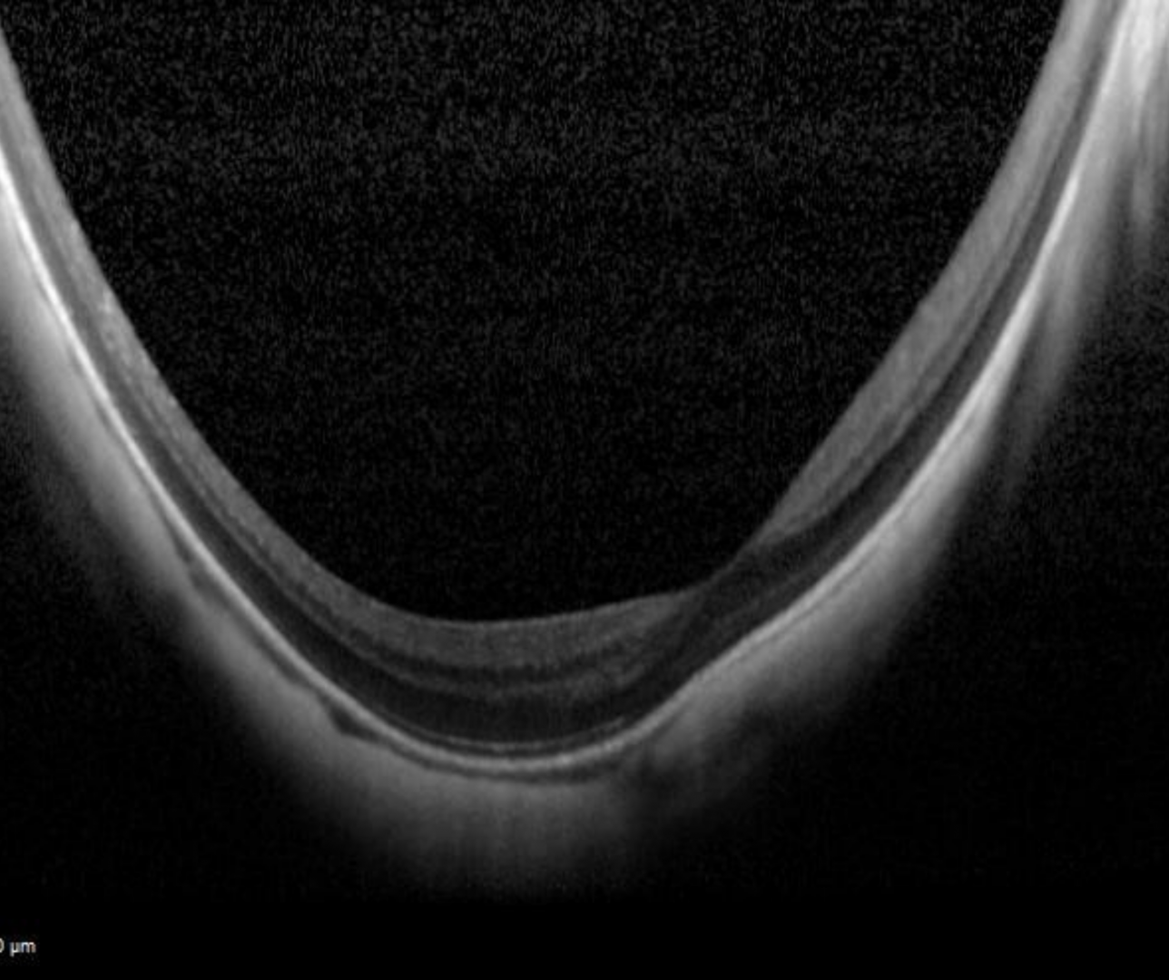
OCT imaging shows marked concavity of the sclera, choroid and retina.
B-scan ultrasound shows a pronounced outpouching caused by the staphyloma.
Case Examples
-
Case 1
A 66 year old Caucasian female.
Right: -1.50 / -5.50 x 125 VA 6/7.5- (20/25-)
Left: -0.75 / -7.00 x 85 VA 6/75+ (20/250+) -
Case 2
A 65 year old Asian male. Cataract was noted in both eyes, more marked in the left eye.
Right: -12.00 / -3.00 x 90 VA 6/9+ (20/30+)
Left: -17.00 / -1,25 x 125 VA 6/24 (20/80)
Differential Diagnosis
References
Ng, D., Cheung, C., Luk, F. et al. (2016) Advances of optical coherence tomography in myopia and pathologic myopia. Eye 30, 901–916
Yasushi, I. (2017) Overview of the complications of high myopia, Retina: Volume 37 - Issue 12 - p 2347-2351