
Overview
Sub-retinal haemorrhage may be associated with lacquer cracks or choroidal neovascularisation in myopic maculopathy.
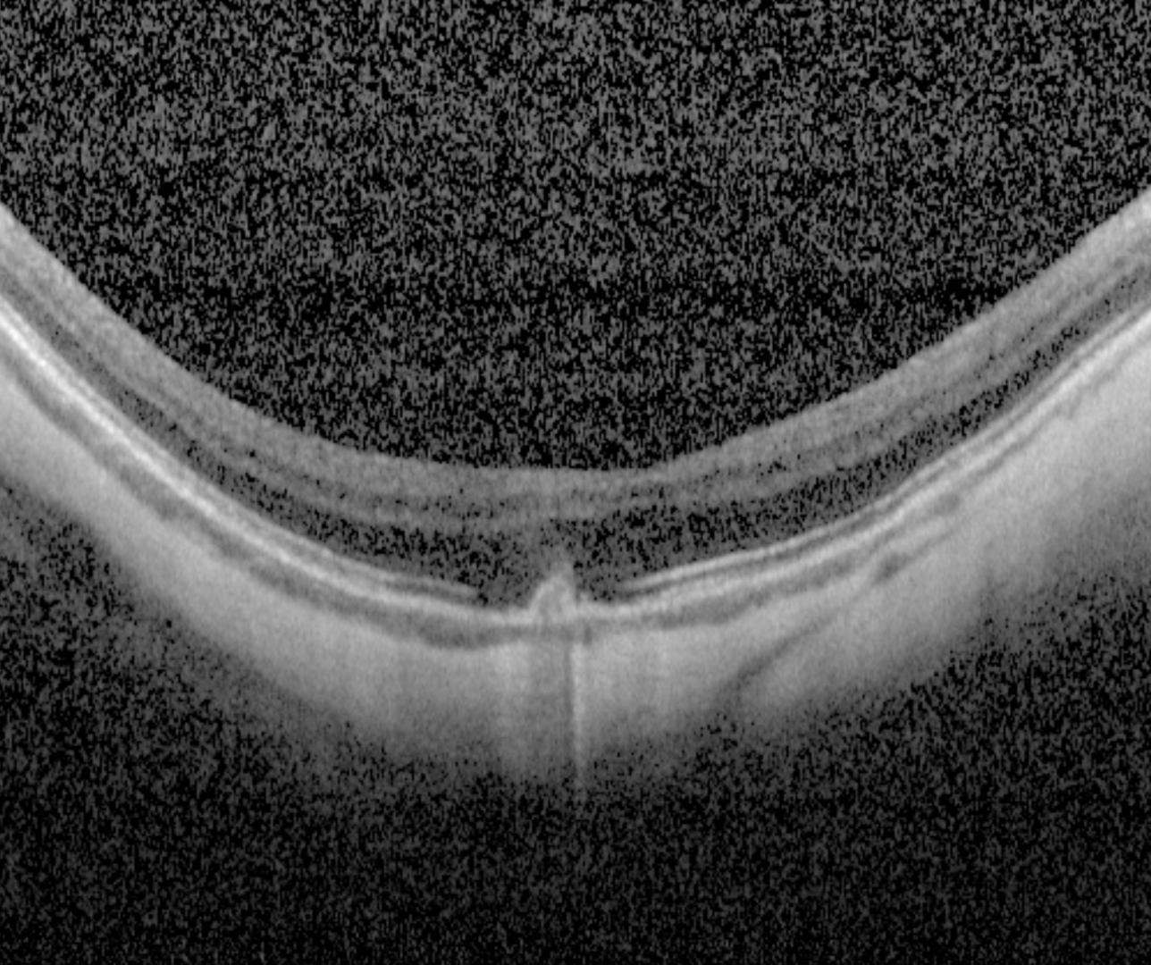
When associated with a lacquer crack, OCT shows a hypo-reflective break in the RPE-Bruch's membrane complex with a "volcanic"-like appearance. There are no signs of associated exudation present.
When associated with myopic CNV, the CNV presents on OCT as a hyper-reflective sub-retinal lesion usually with associated subretinal fluid. More information about myopic CNV is available by clicking on the tab at the bottom of this page.
Case Examples
-
Case 1
A 59 year old Caucasian female with refraction -8.75/-0.5 x 22 and best corrected visual acuity of 6/15 (20/50) in the right eye.
Differential Diagnosis
References
Baba, T. Ohno‐Matsui, K. Yoshida, T. Yasuzumi, K. Futagami, S. Tokoro, T. Mochizuki, M. (2002), Optical coherence tomography of choroidal neovascularization in high myopia. Acta Ophthalmologica Scandinavica, 80:
Hochman MA, Seery CM, Zarbin MA. (1997) Pathophysiology and management of subretinal hemorrhage. Surv Ophthalmol. Nov-Dec;42(3):195-213.