
Overview
The pachychoroid disease spectrum is defined as a group of retinochoroidal diseases unified by distinctive choroidal alterations. Their common imaging characteristics are outlined below.
Conditions that reside within this spectrum include pachychoroid pigment epitheliopathy (PPE), central serous chorioretinopathy (CSCR), pachychoroid neovasculopathy, polypoidal choroidal vasculopathy (PCV), focal choroidal excavation, and peripapillary pachychoroid syndrome (PPS). These conditions are thought to result from similar pathogenesis but represent different clinical manifestations. More information about each can be obtained by clicking on the links at the bottom of this page.
Common Imaging Features
-
Increased choroidal thickness
Subfoveal choroid thickness ranges between 191-350µm (Lehmann, 2015) in normal people.
However, it may be affected by factors such as gender, age, refractive status and even subject to diurnal variations. Choroidal thickness also varies with diseases.
-
Pachyvessels with compression of the overlying choriocapillaris
Dilated choroidal vessels in Haller's layer (known as pachyvessels) are characteristic of this spectrum of diseases. Presence of these pachy vessels are associated with compression of the overlying choriocapillaris as can be seen in the images shown here.
-
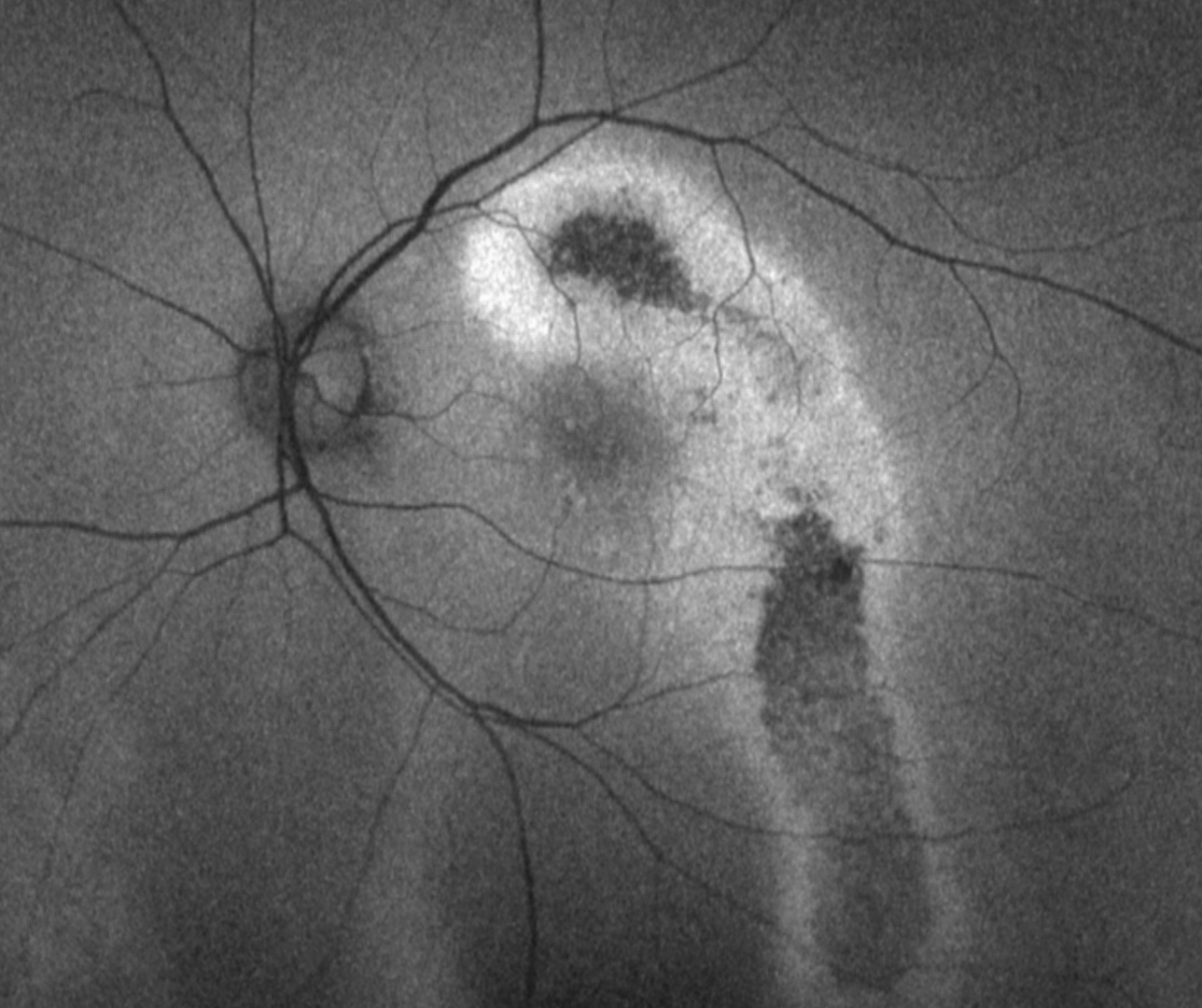
RPE disturbances
RPE disturbances are characteristic of the pachychoroid disease. These disturbances typically overlie an attenuated choriocapillaris and vary in size and appearance with the stage of disease present.
Sub Topics
Pachychoroid pigment epitheliopathy (PPE)
Central serous chorioretinopathy (acute)
Central serous chorioretinopathy (chronic)
Pachychoroid neovasculopathy
Polypoidal choroidal vasculopathy
Focal choroidal excavation
Peripapillary pachychoroid syndrome (PPS)
Peripapillary pachychoroid neovasculopathy (PPN)
References
Borooah, S, Sim, P.Y, Phatak, S, Moraes, G, Wu, C.Y, Cheung, CMG., Pal, B, Bujarborua, D. (2020), Pachychoroid spectrum disease. Acta Ophthalmol.
Cheung, CMG, Lee, WK, Koizumi, H. et al. Pachychoroid disease. Eye 33, 14–33 (2019).