
Overview
Pachychoroid pigment epitheliopathy (PPE) describes pigmentary abnormalities in the context of pachychoroid. The features typical of the pachychoroid disease spectrum are present, including dilated large choroidal vessels with associated attenuation of choriocapillaris and overlying RPE abnormalities. It is generally considered to be a form fruste of central serous chorioretinopathy (CSCR). However, contrary to CSCR, there is no history of or current subretinal fluid present.
Colour fundus photography typically shows mild pigmentary changes and reduced fundus tessellation at the macula and posterior pole.
OCT B-scan shows a range of RPE abnormalities overlying dilated large choroidal vessels. The RPE changes may be drusenoid (termed 'pachydrusen'). Serous pigment epithelial detachment may or may not be present, however, there is no subretinal fluid in PPE.
En face OCT imaging shows dilated large choroidal vessels which do not taper towards the posterior pole and instead maintain their caliber and end abruptly. These dilated or "pachy" vessels can be diffuse or focal.
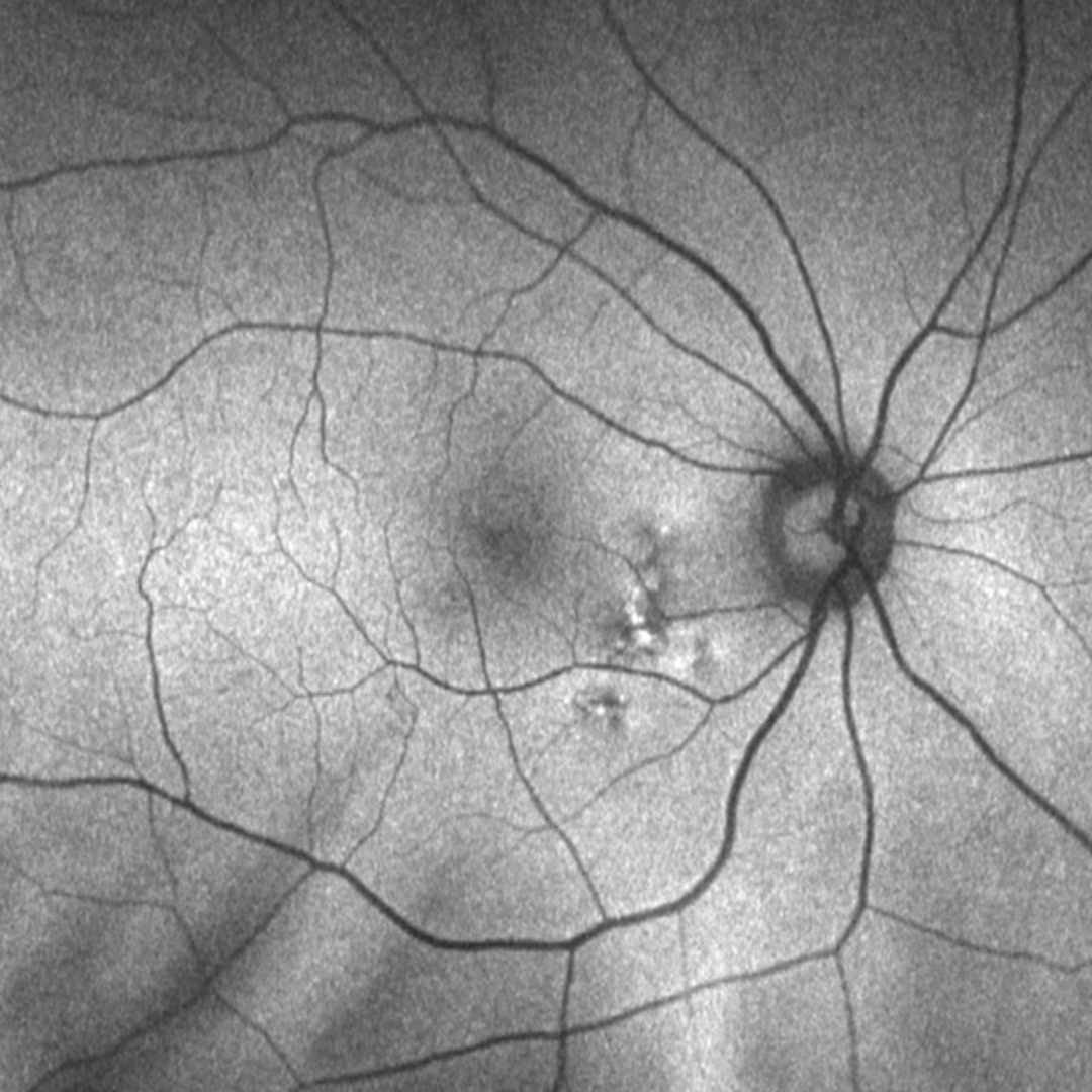
Fundus autofluorescence imaging can show a granular hypo-autoflurorescence or a mixed stippled hypo and/or hyper autofluorescence.
Case Examples
-
Case 1
A 63-year-old Asian female with best-corrected visual acuity of 6/6 (20/20) in each eye.
Fundus photograph and red-free image (right eye)
More infoFundus photograph and red-free image (left eye)
More infoFundus autofluorescence (1-right,2-left) and en face OCT (3-right, 4-left) images
More infoSpectralis OCT volume scans (right and left macula)
More infoSpectralis OCT line scans (left eye)
More info -
Case 2
A 40-year-old Caucasian male with best-corrected visual acuity of 6/6+ (20/20+) in each eye.
References
Borooah, S, Sim, P.Y, Phatak, S, Moraes, G, Wu, C.Y, Cheung, CMG., Pal, B, Bujarborua, D. (2020), Pachychoroid spectrum disease. Acta Ophthalmol.
Cheung, CMG, Lee, WK, Koizumi, H. et al. Pachychoroid disease. Eye 33, 14–33 (2019).