
Peripapillary Pachychoroid Neovasculopathy (PPN)
Peripapillary pachychoroid neovasculopathy (PPN) was only identified in recent years, The term describes peripapillary pachychoroid syndrome (PPS) with the presence of a choroidal neovascular membrane.
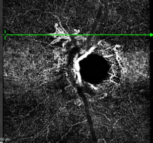
Clinical features include pigmentary changes in the nasal peripapillary area with associated mottled auto-fluorescence. OCT imaging shows dilated "pachy" vessels, the presence of a flat irregular pigment epithelial detachment (FIPED) often with sub or intra-retinal fluid adjacent. OCT angiography shows the choroidal neovascular network in the peripapillary region.
Rare complications can include choroidal folds and optic disc oedema. Referral to an ophthalmologist is required.
Case Examples
-
Case 1: Active PPN
A 70 year old Caucasian male with treated normal tension glaucoma. VA is 6/7.5 in the left eye, not improved by pinhole.
Differential diagnosis
References
B Brown R, Mohan S, Chhablani J. Pachychoroid Spectrum Disorders: An Updated Review. J Ophthalmic Vis Res. 2023 Apr 19;18(2):212-229.
M Hernández J, Remolí Sargues L, Monferrer Adsuara C, Castro Navarro V, Navarro Palop C, Cervera Taulet E. Peripapillary pachychoroid neovasculopathy: A novel entity. Eur J Ophthalmol 2022;32:NP149–NP153