
Overview
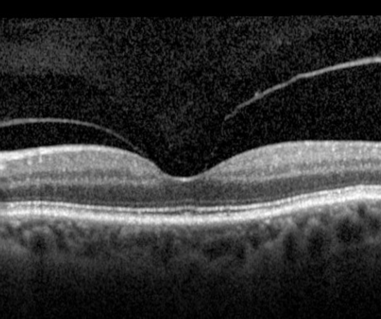
Vitreomacular adhesion (VMA) is defined by perifoveal detachment from the retinal surface but remaining vitreomacular attachment within a 3mm radius of the fovea. There is no detectable distortion of the foveal contour or underlying tissue. Focal adhesion is defined as ≤1500µm where as broad adhesion is defined as >1500µm.
This is a normal finding in the natural course of a PVD.
The macula appearance is unremarkable clinically.
Case Examples
References
Stalmans, P. Duker, J. Kaiser, PK. Heier, JS. Dugel, P et al. (2013) OCT-Based interpretation of the vitreomacular interface and indications for pharmacologic vitreolysis. Retina Volume 33 - Issue 10